Most frequently used radiography is for the periapical which is performed by the bisecting Thus when considering the execution of the radiographic technique and the possibility of errors that occur during the exposure of X-ray image XR receptors it is important to identify those that occur more frequently. The image receptor is placed in a holder and positioned in the mouth parallel to the long axis of the tooth under.

Periapical Radiography Pocket Dentistry
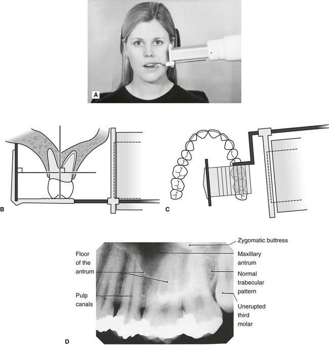
The paralleling technique is recommended for routine periapical radiography but there are.

. The patient is seated upright in the dental chair and should remove any removable dental appliances glasses or jewelry that could interfere with the X-ray beam. Basically there are two techniques for taking periapical radiography. By using a film sensor holder with still.
Paralleling technique Bisecting angle technique Paralleling technique It is also called the extension cone paralleling technique right angle technique and long cone. Demonstration on how to take periapical x-ray using bisecting angle technique. You will need to bite firmly onto the device to keep it in place and provide a clear x-ray image.
These x-rays are often used to detect any unusual changes in the root and surrounding bone structures. In the paralleling technique the central ray of the x-ray beam must be ___ to the film sensor and the long axis of the tooth. Periapical X-rays are used to detect any abnormalities of the root structure and surrounding bone structure.
The X-ray is taken and the exposed plate is then loaded into a scanner or processor which reads the image. This must be done only slightly and the film must never be bent outright. Fitzgerald called as paralleling or long cone technique.
By using a filmsensor holder with fixed image receptor and. Periapical lesions a c ommon dental disease are conventionally detected by dentists through radiog raphy. The film is placed parallel to the long axis of the tooth in question and the central x-ray beam should be directed perpendicular to the long axis of the tooth.
The X-ray tubehead is then aimed at right angles vertically and horizontally to both the tooth and the image. PARALLELING LONG-CONE PERIAPICAL EXPOSURE TECHNIQUES GENERAL A long cone is used to take x-rays with paralleling exposure techniques. The technique involves a constant x-ray cone position which is perpendicular to the floor for maxillary incisors and parallel to the floor for mandibular incisors.
To take a periapical exposure the hygienist or x-ray technician places a small photosensitive imaging plate coated with phosphorus into a sterile wrapper and inserts it into the patients mouth just like a conventional X-ray film card. The periapical film is held between the incisor teeth as if it were an occlusal film for all anterior periapical radiographs. Both techniques have advantages and disadvantages.
The paralleling technique results in good quality x-rays with a minimum of distortion and is the most reliable technique for taking periapical x-rays. How periapical x-rays are taken. Ensure they are seated high enough so it is easy to see the occlusal.
Periapical film is held parallel to the long axis of the tooth using film-holding instruments. The patient was positioned upright with hisher mouth was opened as wide as possible to allow the X-ray beam to pass to the sensor unobstructed from the opposite side of the mouth. A periapical view shows the tooth from the occlusal surface or incisal edge to the.
Gently softening the edges of the film by rounding it very carefully between the fingers. The Bisecting Angle Technique is an alternative to the paralleling technique for taking periapical films. With this technique the film is placed parallel to the long axis of a tooth allowing the X-ray to be focused perpendicular to the long axis of the tooth.
For this purpose a special technique of periapical radiography was developed by Gordon M. The film is placed parallel to the long axis of the tooth in question and the central x-ray beam should be directed perpendicular to the long axis of the tooth. The film is placed parallel to the long axis of the tooth to be radiographed and the central beam of X-ray is directed at right angle to the film and the teeth.
The extraoral periapical radiographic technique was performed for both maxillary and mandibular teeth using Newman and Friedman technique2. The X-ray head is directed at right angles vertically and horizontally of both the tooth and the image receptor. Periapical radiographs provide important information about the teeth and surrounding bone.
Film will be placed near your mouth using a metal rod with a ring attached to it. The paralleling technique results in good quality x-rays with a minimum of distortion and is the most reliable technique for taking periapical x-rays. Periapical radiography is a commonly used intraoral imaging technique in radiology and may be a component of your radiologic examination.
Full Mouth Survey X-rays. The X-ray tube head should be positioned so that the beam meets the tooth and the image. Occlusal X-rays show full tooth development and placement 9.
A periapical x-ray or PA film will show one or two teeth in their entirety in one single image right from the crown of the tooth which is the part exposed in the mouth to the very tips of the tooth roots located in the jawbone as well as. The central ray is directed to pass at a perpendicular angle to both the tooth and the film. This study aimed to assess the application of.
The bisecting short-cone and paralleling long-cone techniques are two of the most commonly used techniques. Extraoral radiograph Panoramic X-ray Tomograms Cephalometric projections Sialography Computed tomography 10. Parallel technique The image receptor is placed in a holder and placed in the mouth parallel to the longitudinal axis of the tooth under.
Single periapical radiographs are often made of individual teeth or groups of teeth to obtain information for treatment or diagnosis of localized diseases or abnormalities. The sensor was placed on the. An intraoral full-mouth survey FMX on an adult consists of _____ images.
Bending the film produces an artifact on the image that may render the film unusable. Periapical X-rays. Some further techniques to make this procedure more comfortable for the patient include.
Bone about 2 to 3 mm beyond the apex. 5 Intra oral Periapical radiography is a commonly used intraoral imaging technique in dental radiology and may be a component of intraoral periapical.
How Make Periapical X Ray
Periapical Radiography Pocket Dentistry

Periapical Radiography Clinical Gate

Periapical Radiography Pocket Dentistry

Periapical Radiography Pocket Dentistry

Periapical Radiography Pocket Dentistry

Periapical Radiography Pocket Dentistry

Periapical Radiography Pocket Dentistry
0 comments
Post a Comment